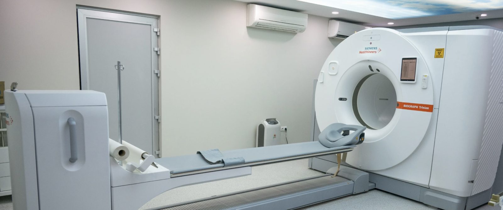
Selçuk University Medical Faculty Hospital’s Nuclear Medicine Clinic has begun using the next-generation digital PET/CT system Biograph Trinion, which is used in the diagnosis and follow-up of a wide range of diseases, particularly cancer. The first and only device of its kind in Konya, it stands out with its high image quality, faster scanning capabilities and artificial intelligence-supported technologies.
The digital PET/CT system added to the Nuclear Medicine Clinic offers significant advantages, particularly in the diagnosis, staging and post-treatment follow-up of cancer patients. Its ultra-fast TOF (Time of Flight) technology enables high-quality images to be obtained in a shorter time. Where appropriate, this can also allow for the use of lower amounts of radioactive tracers and reduce the time patients spend in the scanner. AI-supported imaging technologies further contribute to improved image quality. The scanner’s patient table, which can support patients weighing up to 272 kilograms, will also make imaging services more accessible and comfortable for patients with different physical characteristics.
Selçuk University Rector Prof. Dr. Hüseyin Yılmaz said Biograph Trinion would make a significant contribution to PSMA PET/CT imaging, which is particularly used in the diagnosis and follow-up of prostate cancer.
“Thanks to its high image quality, the system will support the evaluation of small lesions and areas showing low levels of radioactive tracer uptake. With this device, the first and only one of its kind in Konya, we aim to strengthen access to advanced PET/CT services for patients in our region and contribute particularly to the diagnosis and follow-up of cancer patients. We will continue to improve the technological infrastructure of Selçuk University Medical Faculty Hospital in the field of healthcare.”